Friday 5 December 2014

CSEC - Movement of substances

Movement of Substances


Here are the links to see an animation of each movement process discussed in class.

Things to know before we begin


  1. Substances move throughout, in and out of cells by passive and active media. 
  2. A concentration gradient is required to direct the movement
  3. Substances moved are varied and they include small, large, charged and neutral particles
Lets put all this together with a concept map                                                                                    

Now for the details:


  • concentration gradient exists when molecules are not evenly distributed. that means you will have a region having more molecules than another. This difference in the amount of molecules is the concentration gradient.                                                    
  • Movement along a concentration gradient - from an area of high concentration to an area of low concentration.
  • Movement against a concentration gradient - from an area of low concentration to an area of high concentration.
  • Passive processes do not require energy. These include:
    • Diffusion - 
    • Osmosis
  • Active processes require ATP energy.
    • active transport 

  1. Diffusion - movement of uncharged molecules along a concentration gradient, until evenly distributed. eg. perfume in air, red dye in water, oxygen in cells.                   CLICK HERE to see diffusion                                                                                              
  2. Osmosis - movement of water molecules through a selectively permeable membrane, along a concentration gradient, until evenly distributed.                                      CLICK HERE to see osmosis                                                                                            
  3. Active Transport - movement of charged particles (ions) against a concentration gradient through carrier proteins (gates) with the help of energy.                             CLICK HERE to see active transport                                                                             
  4. Endocytosis - the intake of substances into a cell by the folding of the membrane (invagination) into a vacuole.                                                                             > phagocytosis - intake of solid material into a cell                                             >pinocytosis - intake of liquid material into the cell                                                                                                                               
  5. Exocytosis - the release of the contents of a cell vacuole to the outside of the cell by fusion of the vacuole with the cell membrane.                                                         CLICK HERE to see endocytosis & exocytosis 

Friday 28 November 2014

CAPE 1: ENZYMES IN ANIMATION

​Good Morning All, 

I hope these help​....see links below


  • note the key terms and the concepts being explained
  • do NOT click next until the menu bar at the bottom of the page changes its colour from grey to yellow

  • note the difference with how they work
  • select narration
  • read conclusion
  • select home button at the bottom of the animation
  • click step by step and go back through the animation
  • select the "Q" from the bottom of the animation to complete quiz

  • pay attention to what happens to the secondary and tertiary arrangement of the protein
  • most non-specific inhibitors work in this way


Wednesday 26 November 2014

CSEC: CELLS - simple but important.......LEARN IT!

Parts of the UNSPECIALISED cell


"It is important that you learn to DRAW & EXPLAIN the function of these cell organelles" 

Circles and dots will not work when you are representing cell organelles...... 


DIAGRAM OF AN UNSPECIALISED PLANT CELL


Cell Membrane This is a thin, flexible layer round the outside of all cells made of phospholipids and proteins. The function of the cell membrane is to allow waste material to exit the cell. The cell membrane forms a barrier between the inside of the cell and the outside, so that the chemical environments on both sides can be different. It regulates the movement of materials into and out of the cell.


Cell Wall The function of the cell wall is to provide structural support. It gives rigid support due to its strong cellulose component. It has plasmodesmata which are small breaks in the wall itself. They allow substances to enter and leave the cell.


Golgi Body The Golgi body stores, packages, and distributes the lipids and proteins made in the endoplasmic reticulum. It puts proteins into packages, called vesicles.


Rough Endoplasmic Reticulum (RER) The RER is generally a series of connected flattened sacs, studded with numerous ribosomes, which give it its rough appearance. The ribosomes synthesise proteins, which are processed in the RER 


Smooth Endoplasmic Reticulum (SER) Makes lipids and steroids and package proteins for transport.

Lysosomes These are small membrane-bound vesicles formed from the RER containing a mixture of digestive enzymes. They are used to break down unwanted chemicals, toxins, organelles or even whole cells, so that the materials may be recycled. They can also fuse with a feeding vacuole to digest its contents. Lysosomes are the cell’s garbage disposal system. They clean up while patrolling the cell. This organelle breaks down large molecules into many smaller molecules by using their special proteins. Lysosomes also kill and digest invading organisms.


Cytoplasm The function of the cytoplasm is to distribute oxygen and food (nutrients) to other parts of the cell and it supports all parts inside the cell. It has three main functions: storage, energy, and manufacturing. The cytoplasm contains nutrients that have been dissolved which help for the dissolving of waste products.


DIAGRAM OF AN UNSPECIALISED ANIMAL CELL


Vacuole These are membrane-bound sacs containing water or dilute solutions of salts and other solutes. Most cells can have small vacuoles that are formed as required, but plant cells usually have one very large permanent vacuole that fills most of the cell, so that the cytoplasm (and everything else) forms a thin layer round the outside. Plant cell vacuoles are filled with cell sap, and are very important in keeping the cell rigid, or turgid. Some unicellular protoctists have feeding vacuoles for digesting food, or contractile vacuoles for expelling water.

Ribosomes - Ribosomes are the protein builders or protein synthesizers of the cell.


Chloroplast Chloroplasts are the food producers of the cell. They contain chlorophyll, the green pigment that is needed for photosynthesis. Through the process of photosynthesis, green plants use the energy from the sun to convert it into sugar. The main purpose of this organelle is to produce sugars and starches.


Nucleus This is the largest organelle. Surrounded by a nuclear envelope, which is a double membrane with nuclear pores - large holes containing proteins that control the exit of genetic substances and ribosomes from the nucleus. The interior is called the nucleoplasm, which is full of chromatin- a DNA/protein complex. During cell division the chromatin becomes condensed into discrete observable chromosomes.


Nuclear Envelope The nuclear envelope (double membrane) surrounds the nucleus and all of its contents. It is similar to the cell membrane around the whole cell. There are pores and spaces for RNA (genetic materials) and proteins to pass through while the nuclear envelope keeps all of the chromatin and the nucleolus inside.


Nucleolus The nucleolus is where ribosomes are made. The nucleolus disappears during cell reproduction. This is because ribosomes are not needed when cells reproduce.


Mitochondria The function of the mitochondria is to provide the cell with energy. Through the process of respiration, the mitochondria uses oxygen to change sugar into energy. It gives out energy by combining sugar molecules with oxygen to make carbon dioxide and water.

Friday 21 November 2014

CAPE 1: Enzymes and Enzyme Activity

What are enzymes?

Enzymes are globular proteins (biological catalysts). They speed up catalyse) chemical reactions in all living things, and allow them to occur more easily.
They are too small to be seen either when they are inside cells or after they have been released from them, for example in the digestive system.
Each particular enzyme has a unique, 3-dimensional shape shared by all its molecules. Within this shape there is an area called the active site where the chemical reactions occur. 
The active site makes an enzyme specific as it fits only one type of substrate.

How do enzymes work?

Enzymes work by 2 mechanisms:
1. The Lock and Key Model 
    --The enzyme is like a lock with a specific shape into which the key (substrate) fits.
    --Enzymes are usually larger than the substrates that they act on.
     --Once formed, the products cannot fit into the active site and are thus released, leaving the site free.

Click the link to see: the lock and key model in action

2. Induce Fit Model
     --The active site is not rigid and there is no exact fit, instead it can be modified as the substrate interacts with it.
      --The active site is moulded into the shape of the substrate on contact, improving the fit (makes a tighter fit).
Click the link to see: the induce fit model in action


What do enzymes do?

Enzymes lowers the amount of energy required for a chemical reaction to take place. (This energy is called the activation energy.) This causes a reaction involving enzymes to speed up in other words it takes a shorter time for this reaction to form products.

Some enzymes help to break down large molecules. Others build up large molecules from small ones. While many others help turn one molecule into another.

Properties of Enzymes ?

  • Enzymes are catalysts → speed up chemical reactions
  • Reduce activation energy required to start a reaction between molecules
  • Substrates (reactants) are converted into products
  • Reaction may not take place in absence of enzymes (each enzyme has a specific catalytic action)
  • Enzymes remain unchanged at the end of a reaction. [E + S → ES → P + E]
  • Enzymes are specific 

Enzyme activity is  how fast an enzyme is working and is also called the "Rate of Reaction". It is affected by the following factors Temperature, pH, substrate concentration and enzyme concentration.
Although they can do fantastic things they are sensitive and work best under specific conditions.
Each type of enzyme has its own specific optimum conditions under which it works best.
Enzymes work best when they have a high enough substrate concentration for the reaction they catalyse. If too little substrate is available the rate of the reaction is slowed and cannot increase any further.
Copyright S-cool
Sometimes, if too much product accumulates, the reaction can also be slowed down. So it is important that the product is removed.
The pH must be correct for each enzyme. If the conditions are too alkaline or acidic then the activity of the enzyme is affected (it slows down). This happens because the enzyme's shape, especially the active site, is changed. It is denatured, and cannot hold the substrate molecule.
Copyright S-cool
Graph of enzyme activity verses temperature
As the temperature rises, reacting molecules have more and more kinetic energy. This increases the chances of a successful collision and so the rate increases. There is a certain temperature at which an enzyme's catalytic activity is at its greatest (see graph above). This optimal temperature is usually around human body temperature (37.5 oC) for the enzymes in human cells.
Above this temperature the enzyme structure begins to break down (denature) since at higher temperatures intra- and intermolecular bonds are broken as the enzyme molecules gain even more kinetic energy. At very low temperature enzymes are inactive.

Concentration of enzyme and substrate

Graph of enzyme activity verses enzyme concentration           Graph of enzyme activity verses substrate concentration
Graph of enzyme activity verses enzyme concentration           Graph of enzyme activity verses substrate concentration

The rate of an enzyme-catalysed reaction depends on the concentrations of enzyme and substrate. As the concentration of either is increased the rate of reaction increases (see graphs).
For a given enzyme concentration, the rate of reaction increases with increasing substrate concentration up to a point, above which any further increase in substrate concentration produces no significant change in reaction rateThis is because the active sites of the enzyme molecules at any given moment are virtually saturated (occupied) with substrate. The enzyme/substrate complex has to dissociate before the active sites are free to accommodate more substrate. (See graph above on the right)
Provided that the substrate concentration is high and that temperature and pH are kept constant, the rate of reaction is proportional to the enzyme concentration. (See graph above on the left).

Inhibition of Enzyme Activity

Some substances reduce or even stop the catalytic activity of enzymes in biochemical reactions. They block or distort the active site. These chemicals are called inhibitors, because they inhibit reaction.

Inhibitors that occupy the active site and prevent a substrate molecule from binding to the enzyme are said to be active site-directed (or competitive, as they 'compete' with the substrate for the active site).


Inhibitors that attach to other parts of the enzyme molecule, perhaps distorting its shape, are said to be non-active site-directed (or non competitive).


  • All enzymes are globular proteins and round in shape
  • They have the suffix "-ase"
  • Intracellular enzymes are found inside the cell
  • Extracellular enzymes act outside the cell (e.g. digestive enzymes)
  • Enzymes are catalysts → speed up chemical reactions
    • Reduce activation energy required to start a reaction between molecules
    • Substrates (reactants) are converted into products
    • Reaction may not take place in absence of enzymes (each enzyme has a specific catalytic action)
    • Enzymes catalyse a reaction at max. rate at an optimum state
  • Induced fit theory
    • Enzyme's shape changes when substrate binds to active site
    • Amino acids are moulded into a precise form to perform catalytic reaction effectively
    • Enzyme wraps around substrate to distort it
    • Forms an enzyme-substrate complex → fast reaction
    • E + S → ES → P + E
  • Enzyme is not used up in the reaction (unlike substrates)

Changes in pH

  • Affect attraction between substrate and enzyme and therefore efficiency of conversion process
  • Ionic bonds can break and change shape / enzyme is denatured
  • Charges on amino acids can change, ES complex cannot form
  • Optimum pH
    • pH 7 for intracellular enzymes
    • Acidic range (pH 1-6) in the stomach for digestive enzymes (pepsin)
    • Alkaline range (pH 8-14) in oral cavities (amylase)
  • pH measures the conc. of H+ ions - higher conc. will give a lower pH
Enzyme Conc. is proportional to rate of reaction, provided other conditions are constant. Straight line
Substrate Conc. is proportional to rate of reaction until there are more substrates than enzymes present. Curve becomes constant.

Increased Temperature

  • Increases speed of molecular movement → chances of molecular collisions → more ES complexes
  • At 0-42 °C rate of reaction is proportional to temp
  • Enzymes have optimum temp. for their action (varies between different enzymes)
  • Above ≈42°C, enzyme is denatured due to heavy vibration that break -H bonds
    • Shape is changed / active site can't be used anymore

Decreased Temperature

  • Enzymes become less and less active, due to reductions in speed of molecular movement
  • Below freezing point
    • Inactivated, not denatured
    • Regain their function when returning to normal temperature
  • Thermophilic: heat-loving
  • Hyperthermophilic: organisms are not able to grow below +70°C
  • Psychrophiles: cold-loving

Inhibitors

  • Slow down rate of reaction of enzyme when necessary (e.g. when temp is too high)
  • Molecule present in highest conc. is most likely to form an ES-complex
  • Competitive Inhibitors
    • Compete with substrate for active site
    • Shape similar to substrates / prevents access when bonded
    • Can slow down a metabolic pathway
  • [EXAMPLE] Methanol Poisoning
    • Methanol CH3OH is a competitive inhibitor
    • CH3OH can bind to dehydrogenase whose true substrate is C2H5OH
    • A person who has accidentally swallowed methanol is treated by being given large doses of C2H5OH
    • C2H5OH competes with CH3OH for the active site
  • Non-competitive Inhibitors
    • Chemical does not have to resemble the substrate
    • Binds to enzyme other than at active site
    • This changes the enzyme's active site and prevents access to it
  • Irreversible Inhibition
    • Chemical permanently binds to the enzyme or massively denatures the enzyme
    • Nerve gas permanently blocks pathways involved in nerve message transmission, resulting in death
    • Penicillin, the first of "wonder drug" antibiotics, permanently blocks pathways certain bacteria use to assemble their cell wall component (peptidoglycan)

End-product inhibition

  • Metabolic reactions are multi-stepped, each controlled by a single enzyme
  • End-products accumulate within the cell and stop the reaction when sufficient product is made
  • This is achieved by non-competitive inhibition by the end-product
  • The enzyme early in the reaction pathway is inhibited by the end-product
The metabolic pathway contains a series of individual chemical reactions that combine to perform one or more important functions. The product of one reaction in a pathway serves as the substrate for the following reaction.


Curves for reaction rates against substrate concentrations & How to read them

This graph is showing how substrate concentration affects the rate of reaction which is also the speed or velocity of the reaction. The maximum velocity is called Vmax.


The graph levels off at a maximum reaction rate of Vmax. This happens when all the enzyme molecules are working as fast as they can. Once that happens, increasing the concentration of the substrate can't make the reaction go any faster.

What happens to the graph in the presence of a competitive inhibitor?


At a relatively low substrate concentration, the rate of the reaction will be less in the presence of a competitive inhibitor. The competitive inhibitor is just getting in the way of the substrate by attaching itself to some of the active sites.

However, as the substrate concentration increases, the substrate out-competes the inhibitor. If there is a lot more substrate than inhibitor, the chances are far higher that a substrate molecule will hit an active site than an inhibitor molecule will.

That means that at a high enough substrate concentration, the maximum reaction rate will again be Vmax.


What happens to the graph in the presence of a non-competitive inhibitor?


This is quite different.

Once a non-competitive inhibitor has attached itself to the enzyme, that particular enzyme molecule won't work any more. The attachment may be reversible, but even if it is, there will be a proportion of the enzyme molecules which are out of action at any one time.

That means that however much you may increase the concentration of the substrate, you will never reach the original Vmax.

So a graph involving a non-competitive inhibitor looks like this:


There is now a new Vmax, lower than the one where there was no inhibitor present.


Combining these two graphs


In order to get the full mark for the non-competitive line, it had to start off more steeply than the other one, and then it may or may not cross - as I have shown in this diagram. This is so because "The initial rate of a non-competitively inhibited reaction could be greater or less than that of a competitively inhibited reaction. If greater, the curves will cross; if less, they won't cross.”

Tuesday 28 October 2014

CSEC: Classifying Animals

See animations below:

  • click "play movie" which will take you through the distinguishing feature between the different phylum

  • click on each word to obtain a definition....this is just to make sure

  • Let's see if you learnt anything with this EASY quiz
  • Click online quiz
  • select correct answers

  • So how about trying the hard quiz?
HAVE FUN!!!!!!

CAPE 2: Nitrogen Cycle


see animations below: 

  • learn the names of each step and what happens here

  • note what happens in each step
Now practice drawing the cycle on your own

How did you with this simple version? 

Let's try  drawing option number 2
How did you do with this one?

Let's wrap it up with a complete cycle


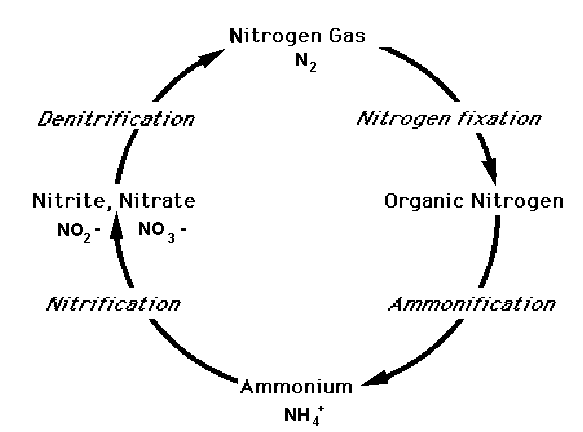
Microbes are involved at most stages of the nitrogen cycle:

Nitrogen Fixation. 78% of the atmosphere is nitrogen gas (N2), but this is inert and can’t be used by plants or animals. Nitrogen fixing bacteria reduce nitrogen gas to ammonia (N2 + 6H g 2NH3), which dissolves to form ammonium ions (NH4+ ). This process uses the enzyme nitrogenase and ATP as a source of energy. The nitrogen-fixing bacteria may be free-living in soil or water, or they may live in colonies inside the cells of root nodules of leguminous plants such as clover or peas. This is an example of mutualism as the plants gain a source of useful nitrogen from the bacteria, while the bacteria gain carbohydrates and protection from the plants. Nitrogen gas can also be fixed to ammonia by humans using the Haber process, and a small amount of nitrogen is fixed to nitrate by lightning.

Nitrification. Nitrifying bacteria can oxidise ammonia to nitrate in two stages: first forming nitrite ions NH4+gNO-2  then forming nitrate ions NO-2gNO-3. These are chemosynthetic bacteria, which means they use the energy released by nitrification to live, instead of using respiration. Plants can only take up nitrogen in the form of nitrate.

Denitrification. The anaerobic denitrifying bacteria convert nitrate to N2 and NOx, which is then lost to the air. This represents a constant loss of “useful” nitrogen from soil, and explains why nitrogen fixation by the nitrifying bacteria and fertilisers are so important.

Ammonification. Microbial saprophytes break down proteins in detritus to form ammonia in two stages: first they digest proteins to amino acids using extracellular protease enzymes, then they remove the amino groups from amino acids using deaminase enzymes.


Monday 27 October 2014

CAPE 1: CELL STRUCTURE & CELL MEMBRANE

Here are animations to help you grasp the concepts:

  • pass mouse over cell organelles to view text on functions
  • right click on the golgi apparatus and see how it works

  • click play

  • read text and play each animation

  • calculating water potential

  • factors affecting water potential 

  • concept as you know it, look out for term concentration gradient 

  • review the concept of osmosis as you know it with the introduction of of the term osmotic concentration

  • look out of key concepts such as: hypertonic, hypotonic, plasmolysis

  • introduction of term protein carrier

  • note the role of ATP in the process
  • note the role of the carrier proteins
  • not the ratio and movement of sodium and potassium ions
  • note change in conformation of carrier proteins

  • cement concept learnt in previous video

P.S. WORK ON THE MCQ QUIZ BELOW EACH VIDEO 

Wednesday 22 October 2014

Drawing Rules for Biological Drawings

Drawing Rules:

• Drawings should be done on plain paper (without lines) only.

• Paper should be bordered 1 cm form the margin at all for sides.

• Only sharp pencils should be used on your drawing paper (NO PEN).

• Each drawing should have a title which is to be written in CAPITAL letters and can be place at the bottom of the paper
It should begin as follows eg. DRAWING OF A _______________.

• Each drawing must have a magnification; this will give you the comparative size of your drawing to that of the original specimen. It can be calculated as follows
Magnification = length of drawing / length of specimen
The figure is always to 2 decimal places. Eg. Mg X 0.53 (no units required)

• Both the title and magnification should be singly underlined with the exception of Scientific names which will double underlined and written using the rules for written scientific names.

• Drawings should be large, clean and clear and to one side of the paper this is to accommodate labels.

• Smooth continuous lines should be use, no sketching or double lines allowed.


• Label lines should be drawn with a straight edge and should be parallel to each other (they should never cross), they should all end at the same place on the page.


• Labels should be to one side of the drawing (no arrow heads at the end if label lines)

• Labels should be written with pencil in lower case script

• No shading allowed instead you are allowed to stipple (use dots) or dashes (-) or lines to show contrast

Thanks to drawitneat for the diagrams

Rules for drawing graphs

Rules for drawing graphs 
 Ø  Graphs must be done on graph paper using only a pencil.

 Ø  All writing on graph paper must be done in script.

 Ø  Pencil should be pointed so that lines drawn are thin and accurately placed.

 Ø  All graphs must have a title eg. Bar graph showing________________

 Ø  Each axis must be appropriately labelled with graduations, what these graduations represent  as well as the related units.

 Ø  The scale of each axis must be clearly placed on the graph paper.

 Ø  When needed symbols to be used to identify strategic points must include `+’ (a cross not an` x’), (a dot), (a dot with a surrounding circle)


 Ø  Broken or unbroken lines can be used to differentiate graphs on the same graph paper. 

 Ø  If a key is necessary, one must be included.


When plotting a graph the:-
 independent variable goes on the horizontal axis
 dependent variable goes on the vertical axis