Wednesday, 22 October 2014

Rules for drawing graphs

Rules for drawing graphs 
 Ø  Graphs must be done on graph paper using only a pencil.

 Ø  All writing on graph paper must be done in script.

 Ø  Pencil should be pointed so that lines drawn are thin and accurately placed.

 Ø  All graphs must have a title eg. Bar graph showing________________

 Ø  Each axis must be appropriately labelled with graduations, what these graduations represent  as well as the related units.

 Ø  The scale of each axis must be clearly placed on the graph paper.

 Ø  When needed symbols to be used to identify strategic points must include `+’ (a cross not an` x’),● (a dot), (a dot with a surrounding circle)


 Ø  Broken or unbroken lines can be used to differentiate graphs on the same graph paper. 

 Ø  If a key is necessary, one must be included.


When plotting a graph the:-
 independent variable goes on the horizontal axis
 dependent variable goes on the vertical axis


Tuesday, 14 October 2014

CAPE 1 - CELLS and CELL STRUCTURE

When you look at animal or plant cells under the electron microscope, you can see a lot more detail. You are able to see the inside structures – organelles – of the cells, which together make a cell’s ultrastructure. Most organelles are common to both animal and plant cells. They have the same function in teach type of cell. Each organelle has its own specific role within the cell, all working together and each contributing towards the survival of the cell. This process is called division of labour.


CYTOSKELETON 

Cells contain a network of fibres made of protein. These fibres keep the cell’s shape stable by providing an internal framework called the cytoskeleton:
-->Some of the fibres, called  microfilaments (made of actin filaments) are able to move against each other – these cause the movement seen in some white blood cells, and they move some organelles around inside cells.  Movement is side-to-side like a wind-shield wiper.
-->There are other fibres, called microtubules. These are cylinders about 25nm in diameter made of a protein called tubulin, and may be used to move a microorganism through a liquid or to waft a liquid past a cell. Movement is circular like a helicopter propeller.

Comparing Micortubule & Microfilaments:

Arrangement of microtubules- arranged in a 9+2 arrangement seen below:


UNDULIPODIA & CILIA

Structurally, flagella of eukaryotes (correctly named undulipodia) and cilia are the same. Each one is made up of a cylinder than contains nine microtubules arranged in a circle and another two microtubules in a central bundle. Undulipodia are longer than cilia.
The undulipodium that forms the tail of a sperm cell can move the entire cell. Undulipodia and cilia can move because the microtubules can use energy produced by ATP (adenosine triphosphate).
Some bacteria have flagella. These look like the same as eukaryotic undulipodia, but their internal structure is different. These are true motors; they are made of a spiral of protein, called flagellin, attached by a hook to a protein disc at the base. Using energy from ATP, the disc rotates, spinning the flagellum
Cell ultrastructure and the importance of the cytoskeleton of cells.
M
any of the organelles found within cells are membrane-bound, this means that they have their own surrounding membranes to separate them from the rest of the contents of the cell. They have the same structure as the main cell membrane. The organelles form separate compartments within the cell, a process called compartmentalisation.
Structure
Function
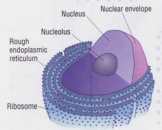
The nucleus is the largest organelle in the cell. When stained, it shows darkened patches known as chromatin. It is surrounded by a nuclear envelope. This is a structure made of two membranes with fluid between them. A lot of holes, called nuclear pores, go right through the envelope. These holes are large enough for relatively large molecules to pass through. There is a dense, spherical structure, called the nucleolus, inside the nucleus
The nucleus stores the majority of the cell’s genetic material. The chromatin consists of DNA and proteins. It contains the instructions for making proteins. Some of these proteins regulate the cell’s activities. When a cell divides, chromatin condenses into visible chromosomes. The nucleolus makes RNA and ribosomes. These pass into the cytoplasm and proteins are assembled at them
Endoplasmic reticulum (ER) consists of a series of flattened membrane-bound sacs called cisternae. They are continuous with the outer nuclear membrane. Rough ER is studded with ribosomes, smooth ER does not have ribosomes
Rough ER transports proteins that were made on the attached ribosomes. Some of these proteins may be secreted from the cell. Some will be placed on the cell surface membrane. Smooth ER is involved in making the lipids that the cell needs
The Golgi apparatus is a stack of membrane-bound sacs, which looks very much like a pitta bread
The Golgi apparatus is responsible for receiving proteins and modifying them. It receives proteins from the ER and may add sugar molecules to them. It then packages the modified proteins into vesicles that can be transported. Some modified proteins go to the cell surface so they can be secreted
A single mitochondrion is spherical or sausage-shaped. It has two membranes separated by a fluid-filled space. The inner membrane is highly-folded to form cristae. The central part of the mitochondrion is the matrix
Mitochondria are the site where ATP is produced during respiration.  ATP is sometimes called the universal carrier energy as it drives most of the cellular processes
Chloroplasts are only found in plant cells, and have two membranes separated by a fluid-filled space. The inner membrane is continuous, with an elaborate network of flattened membrane sacs called thylakoids. A stack of thylakoids is a granum (plural: grana). Chlorophyll molecules are present on the thylakoids membranes and in the intergranal membranes
These are the site of photosynthesis in plant cells. Light energy is used to drive the reactions, in which carbohydrate molecules are made from carbon dioxide and water
A lysosome is a spherical sac surrounded by a single membrane
These contain powerful digestive enzymes which are there to break down materials. For example, white blood cell lysosomes help to break down invading microorganisms; and the specialised lysosome in the head of a sperm cell helps penetrate the female egg cell

►Golgi apparatus


                                                                                                              ◄The nucleus and endoplasmic reticulum


                                                                                     ◄Mitochondrion


               ► Chloroplast
 

There are some organelles which are non membrane-bound…
Structure
Function
A ribosome is a tiny organelle that consists of two subunits. They can be found in the cytoplasm or attached to the ER making rough ER
Ribosomes are the site of protein synthesis in the cell (where new proteins are made). They act as an assembly line where coded information (mRNA) from the nucleus is used to assemble proteins from amino acids
Centrioles are small tubes of protein fibres (microtubules) which are present only in animal cells and cells of some protoctists. They are found in a pair next to the nucleus
These are used in cell division, they form fibres known as spindle which move the chromosomes during nuclear division


CAPE 2 & CSEC - Nutrient Cycles - Carbon & Nitrogen cycle

Nutrient Cycles

  • Elements are taken up by producers (plants) / stored as organic matter
  • Passed on across trophic level / consumer digest and absorb food / stored as organic matter
  • Decomposers decay detritus and excretory products / return inorganic ions to environment / taken up by producers
  • Warm temp / higher enzyme activity / faster decomposition
Table 9-14-1: Use of nutrients in plants and animals


PLANTS
ANIMALS
CARBONOrganic substances / lipids /
proteins / ATP / chlorophyll
Organic substances / lipids /
proteins / ATP / chlorophyll
NITROGEN- Amino acid / nucleotide synthesise
- In RNA, DNA, proteins, ATP
- Amino acid / nucleotide synthesise
- In RNA, DNA, proteins, ATP
IRON- In cytochromes / ETC
- Needed for enzymes such as
catalase to work
- Synthesis of chlorophyll
- In cytochromes / ETC
- Needed for enzymes such as
catalase to work
- Part of Hb
IODINEContained in thyroxine (hormone)
MOLYBDENUMNitrate reductase / reduces nitrates during synthesis of amino acids

Carbon Cycle

  • Producers, consumers, decomposers
    • Add CO2 to the air by respiration
    • Carbon is stored in tissues as organic matter (carbohydrates, lipids, proteins)
    • Carbon is passed along food web by feeding
  • Plants remove CO2 from air by photosynthesis
  • Animals excrete carbon as waste products
  • Decomposers decay detritus and excretory products / add carbon to soil
    • Detrivores digest detritus to small pieces / large surface area
    • Saprophytes digest smaller detritus by
      • Extracellular digestion by secreting enzymes
      • Absorb resulting nutrients across plasma membrane
      • Releases inorganic matter (CO2, H2O, mineral ions) into soil
  • Fossil fuels
    • Combustion releases CO2 into air
    • Fossilisation of carbon atoms in organic compounds in dead remains (plants, animals) and excretory products (animals)
  • Respiring organisms must not die to release stored carbon / differs from other cycles

Respiration, Photosynthesis and CO2

  • Photosynthesis takes up more CO2 than is released by respiration
  • CO2 concentration
    • Higher at night than at daylight; light-dependent reaction cannot take place
    • Peaks at winter time due to high oil consumption; low rate of photosynthesis due to cooler temp, shorter day length, loss of leaves
  • Variation in a graph due to wind mixing CO2 with the surrounding air
    • Graph should show conc over whole area rather over a specific area
  • Rate of photosynthesis and respiration are balanced in a rain forest
    • Forests grow for a long time and have stored lots of carbon in their tissues, other plants have stored carbon as cellulose and lignin

Nitrogen Cycle

  • *processes involved in restoring nitrate conc in soil after cultivation is abandoned

1) Assimilation (→Building up organic molecules)

  • Plants take up NITRATE NO3/AMMONIA NH3 from the soil by active transport
    • Used to synthesis amino acids / synthesise proteins / new cells and tissues
  • Primary consumers feed on plants
    • Proteins are digested into amino acids and absorbed
    • Amino acids synthesise new proteins
  • Nitrogen is passed along the trophic level through the food web

2) Ammonification*

  • Detritus/leaves from plants/excretion from animals/dead animals
  • Broken down by saprotrophs/decomposition
  • Releases ammonia (NH3)/ammonium ions (from decay)
  • Ammonia dissolves in H2O → NH3 + aq → NH4+

3) Nitrification*

  • Ammonium NH4+ / nitrite NO2-
  • Nitrite / nitrate
  • By aerobic nitrifying bacteria eg. include: 
    • (Nitrosomonas, Nitrosospira, Nitrosococcus, and Nitrosolobus - which are bacteria that convert ammonia to nitrites), 
    • (Nitrobacter,Nitrospina, and Nitrococcus - which are bacteria that convert nitrites (toxic to plants) to nitrates )

4) Denitrification

  • Removal of nitrogen from NO2 (nitrite)- and NO3 (nitrate)- to make N2(g)
  • By anaerobic denitrifying bacteria eg. include:
    •  Thiobacillus denitrificans, Micrococcus denitrificans, and some species of Serratia, Pseudomonas, and Achromobacter.  

5) Nitrogen Fixation*


  • N2(g) is converted to nitrates by lightning N2(g) + O2 → NO3-
    • NITROGEN GAS IS CONVERTED TO NH3/NH4+
  • By Haber process: N2(g) + H2 → ammonia NH3
    • //used to make fertilisers / added to soil / leakage of ions into river
  • By Nitrogen-fixing bacteria by anaerobic nitrogenase. There are 2 types of nitrogen fixing bacteria:
    • Live free in soil (non-symbiotic) bacteria, including the cyanobacteria (or blue-green algae) Anabaena, Azotobacter, Beijerinckia, and Clostridium;
    •  Mutualistic (symbiotic) bacteria such as Rhizobium, living in the nodules of leguminous plants, and Spirillum lipoferum, associated with cereal grasses.

Tuesday, 30 September 2014

LIPIDS - CAPE U1

Lipids 

Lipids are a mixed group of hydrophobic compounds composed of the elements carbon, hydrogen and oxygen.

Structure:
􀀀glycerol (3C alcohol) + fatty acid
􀀀fatty acid = long HC “tail” with carboxyl (COOH) group “head

Triglycerides 


􀀀3 fatty acids linked to glycerol

􀀀ester linkage = between OH & COOH


Triglycerides are commonly called fats or oils. They are made of glycerol and fatty acids.
Glycerol is a small, 3-carbon molecule with three alcohol groups.
Fatty acids are long molecules with a polar, hydrophilic end and a non-polar, hydrophobic "tail". The hydrocarbon chain can be from 14 to 22 CH2units long, but it is always an even number because of the way fatty acids are made. The hydrocarbon chain is sometimes called an R group, so the formula of a fatty acid can be written as R-COO-.
  • If there are no C=C double bonds in the hydrocarbon chain, then it is a saturated fatty acid (i.e. saturated with hydrogen). These fatty acids form straight chains, and have a high melting point.
  • If there are C=C double bonds in the hydrocarbon chain, then it is an unsaturated fatty acid (i.e. unsaturated with hydrogen). These fatty acids form bent chains, and have a low melting point. Fatty acids with more than one double bond are called poly-unsaturated fatty acids (PUFAs).
One molecule of glycerol joins together with three fatty acid molecules to form a triglyceride molecule, in another condensation polymerisation reaction: see animation formation of triglycerides
Triglycerides are insoluble in water. They are used for storage, insulation and protection in fatty tissue (or adipose tissue) found under the skin (sub-cutaneous) or surrounding organs. They yield more energy per unit mass than other compounds so are good for energy storage. Carbohydrates can be mobilised more quickly, and glycogen is stored in muscles and liver for immediate energy requirements.
  • Triglycerides containing saturated fatty acids have a high melting point and tend to be found in warm-blooded animals. At room temperature thay are solids (fats), e.g. butter, lard.
  • Triglycerides containing unsaturated fatty acids have a low melting point and tend to be found in cold-blooded animals and plants. At room temperature they are liquids (oils), e.g. fish oil, vegetable oils.


Phospholipids

Phospholipids have a similar structure to triglycerides, but with a phosphate group in place of one fatty acid chain. There may also be other groups attached to the phosphate. Phospholipids have a polar hydrophilic "head" (the negatively-charged phosphate group) and two non-polar hydrophobic "tails" (the fatty acid chains). This mixture of properties is fundamental to biology, for phospholipids are the main components of cell membranes.
When mixed with water, phospholipids form droplet spheres with the hydrophilic heads facting the water and the hydrophobic tails facing eachother. This is called a micelle.
 Alternatively, they may form a double-layeredphospholipid bilayer. This traps a compartment of water in the middle separated from the external water by the hydrophobic sphere. This naturally-occurring structure is called a liposome, and is similar to a membrane surrounding a cell.

Waxes

Waxes are formed from fatty acids and long-chain alcohols. They are commonly found wherever waterproofing is needed, such as in leaf cuticles, insect exoskeletons, birds' feathers and mammals' fur.

Steroids

Steroids are small hydrophobic molecules found mainly in animals. They include:
  • cholesterol, which is found in animals cell membranes to increase stiffness
  • bile salts, which help to emulsify dietary fats
  • steroid hormones such as testosterone, oestrogen, progesterone and cortisol
  • vitamin D, which aids Ca2+ uptake by bones.

Terpenes

Terpenes are small hydrophobic molecules found mainly in plants. They include vitamin A, carotene and plant oils such as geraniol, camphor and menthol.

Sunday, 28 September 2014

CAPE 1 - Carbohydrates

Carbohydrates  

Carbohydrates contain only the elements carbon, hydrogen and oxygen. The group includes monomers, dimers and polymers, as shown in this diagram:

Review Table


Monosaccharides (simple sugars) 

These all have the formula (CH2O)n, where n can be 3-7. The most common and important monosaccharide is glucose, which is a six-carbon or hexose sugar, so has the formula C6H12O6. Its structure is:
a-glucose (used to make starch and glycogen)
or more simply
b-glucose (used to make cellulose)

 Glucose forms a six-sided ring, although in three-dimensions it forms a structure that looks a bit like a chair. The six carbon atoms are numbered as shown, so we can refer to individual carbon atoms in the structure. In animals glucose is the main transport sugar in the blood, and its concentration in the blood is carefully controlled. There are many isomers of glucose, with the same chemical formula (C6H12O6), but different structural formulae. These isomers include fructose and galactose.

Common five-carbon, or pentose sugars (where n = 5, C5H10O5) include ribose and deoxyribose (found in nucleic acids and ATP) and ribulose (which occurs in photosynthesis).

Disaccharides (double sugars) 

Disaccharides are formed when two monosaccharides are joined together by a glycosidic bond. The reaction involves the formation of a molecule of water (H2O) and is known as dehydration or condensation: 

see animation: dehydration or condensation of monosaccharides


This shows two glucose molecules joining together to form the disaccharide maltose. Because this bond is between carbon 1 of one molecule and carbon 4 of the other molecule it is called a 1-4 glycosidic bond. Bonds between other carbon atoms are possible, leading to different shapes, and branched chains.
This kind of reaction, where H2O is formed, is called a condensation reaction. 

The reverse process, when bonds are broken by the addition of water (e.g. in digestion), is called a hydrolysis reaction. 

see animation:hydrolysis of carbohydrates (di- and polysaccharides 

In general:
  •  polymerisation reactions are condensations
  • breakdown reactions are hydrolyses
 There are three common disaccharides:
  • Maltose (or malt sugar) is glucose 1-4 glucose. It is formed on digestion of starch by amylase, because this enzyme breaks starch down into two-glucose units. Brewing beer starts with malt, which is a maltose solution made from germinated barley. Maltose is the structure shown above.
  • Sucrose (or cane sugar) is glucose 1-2 fructose. It is common in plants because it is less reactive than glucose, and it is their main transport sugar. It is the common table sugar that you put in your tea.
  • Lactose (or milk sugar) is galactose 1-4 glucose. It is found only in mammalian milk, and is the main source of energy for infant mammals.

Polysaccharides 

Polysaccharides are long chains of many monosaccharides joined together by glycosidic bonds. There are three important polysaccharides:
  • Starch is the plant storage polysaccharide. It is insoluble and forms starch granules inside many plant cells. Being insoluble means starch does not change the water potential of cells, so does not cause the cells to take up water by osmosis (more on osmosis later). It is not a pure substance, but is a mixture of amylose and amylopectin.
Amylose is simply poly-(1-4) glucose, so is a straight chain. In fact the chain is floppy, and it tends to coil up into a helix.
Amylopectin is poly(1-4) glucose with about 4% (1-6) branches. This gives it a more open molecular structure than amylose. Because it has more ends, it can be broken more quickly than amylose by amylase enzymes.

Both amylose and amylopectin are broken down by the enzyme amylase into maltose, though at different rates.
  • Glycogen is similar in structure to amylopectin. It is poly (1-4) glucose with 9% (1-6) branches. It is made by animals as their storage polysaccharide, and is found mainly in muscle and liver. Because it is so highly branched, it can be mobilised (broken down to glucose for energy) very quickly.


  •  Cellulose is only found in plants, where it is the main component of cell walls. It is poly (1-4) glucose, but with a different isomer of glucose. Starch and glycogen contain a-glucose, in which the hydroxyl group on carbon 1 sticks down from the ring, while cellulose contains b-glucose, in which the hydroxyl group on carbon 1 sticks up. This means that in a chain alternate glucose molecules are inverted.
This apparently tiny difference makes a huge difference in structure and properties. While the a1-4 glucose polymer in starch coils up to form granules, the b14 glucose polymer in cellulose forms straight chains. Hundreds of these chains are linked together by hydrogen bonds to form cellulose microfibrils. These microfibrils are very strong and rigid, and give strength to plant cells, and therefore to young plants and also to materials such as paper, cotton and sellotape.
The b-glycosidic bond cannot be broken by amylase, but requires a specific cellulase enzyme. The only organisms that possess a cellulase enzyme are bacteria, so herbivorous animals, like cows and termites whose diet is mainly cellulose, have mutualistic bacteria in their guts so that they can digest cellulose. Humans cannot digest cellulose, and it is referred to as fibre.

Chitin - (poly glucose amine), found in fungal cell walls and the exoskeletons of insects. The structure resembles that of cellulose, except that the hydroxyl groups on C# 2 have been replaced by acetylamino groups. 


  • Other polysaccharides that you may come across include:
  • .Pectin (poly galactose uronate), found in plant cell walls.
  • Agar (poly galactose sulphate), found in algae and used to make agar plates.
  • Murein (a sugar-peptide polymer), found in bacterial cell walls.
  • Lignin (a complex polymer), found in the walls of xylem cells, is the main component of wood.